

© All rights reserved galwayclinic.com 2013


Galway Clinic Facebook page
Tel: +353 (0) 91720170 brendan.ocochlain@galwayclinic.com


Catheter Ablation
Radiofrequency catheter ablation
Radiofrequency cather ablation is a technique used to treat arrhythmias. These are abnormal heart rhythms created by a disturbance in the heart's electrical system. They are usually present from birth and only manifest themselves as patents get older. They usually become more frequent and severe over time. Catheter ablation destroys or disrupts parts of the electrical pathways causing the arrhythmias. With this minimally invasive technique, developed in the 1980s, patients usually leave the hospital in one day with a permanent cure of their arrhythmia. A catheter is placed into a vein in the leg and advanced through the vein to the heart under X ray guidance. Using a cautery tip or radiofrequency energy, the abnormal tissue circuit causing the arrhythmia is cauterised or ablated.The technique
The technique has become the treatment of choice for patients with arrhythmias who do not respond well to drug therapy or who have
a propensity for rapid heart rates. Many patients have side effects with medications which would have to be taken daily for life.
Catheter ablation also is a good option for patients who do not respond well to medication, as well as the elderly who are prone to
suffer side effects from drug therapy. An ablation will lead to a permanent cure for a condition which otherwise would be lifelong.
Recent studies
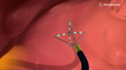
Recent studies of catheter ablation patients with dangerously rapid heart rates found that one month after the procedure, 98 percent required no medication and 95 percent reported considerable improvement in their overall health. In addition, patients reported improvements in their ability to work, exercise and take on physical activities. Procedure During the procedure, a tiny metal-tipped wire catheter is threaded through a vein in the leg up into the heart. Using X ray guidance, the electrophysiologist (specialised cardiologist) can view the catheter moving through the vessel on a monitor. Other catheters containing electrical sensors also are inserted, usually through the leg veins and are used to help find the areas causing the arrhythmia. Once these problem areas are located, the metal-tipped catheter is used to deliver radiofrequency waves — the same energy used for radio and television transmission — to gently burn away the problem tissue. Patients are able to go home the following morning. Many people resume their normal daily activities, such as walking and bathing, upon discharge and return to work in a few days. That said, each patient's experience is slightly different and patients should always follow their doctors instructions.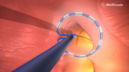


Galway Site Design

Galway Clinic Facebook page


Catheter Ablation
Radiofrequency catheter ablation
Radiofrequency cather ablation is a technique used to treat arrhythmias. These are abnormal heart rhythms created by a disturbance in the heart's electrical system. They are usually present from birth and only manifest themselves as patents get older. They usually become more frequent and severe over time. Catheter ablation destroys or disrupts parts of the electrical pathways causing the arrhythmias. With this minimally invasive technique, developed in the 1980s, patients usually leave the hospital in one day with a permanent cure of their arrhythmia. A catheter is placed into a vein in the leg and advanced through the vein to the heart under X ray guidance. Using a cautery tip or radiofrequency energy, the abnormal tissue circuit causing the arrhythmia is cauterised or ablated.The technique
The technique has become the treatment of choice for patients with
arrhythmias who do not respond well to drug therapy or who have
a propensity for rapid heart rates. Many patients have side effects
with medications which would have to be taken daily for life.
Catheter ablation also is a good option for patients who do not
respond well to medication, as well as the elderly who are prone to
suffer side effects from drug therapy. An ablation will lead to a
permanent cure for a condition which otherwise would be lifelong.
Recent studies
Recent studies of catheter ablation patients with dangerously rapid heart rates found that one month after the procedure, 98 percent required no medication and 95 percent reported considerable improvement in their overall health. In addition, patients reported improvements in their ability to work, exercise and take on physical activities. Procedure During the procedure, a tiny metal-tipped wire catheter is threaded through a vein in the leg up into the heart. Using X ray guidance, the electrophysiologist (specialised cardiologist) can view the catheter moving through the vessel on a monitor. Other catheters containing electrical sensors also are inserted, usually through the leg veins and are used to help find the areas causing the arrhythmia. Once these problem areas are located, the metal-tipped catheter is used to deliver radiofrequency waves — the same energy used for radio and television transmission — to gently burn away the problem tissue. Patients are able to go home the following morning. Many people resume their normal daily activities, such as walking and bathing, upon discharge and return to work in a few days. That said, each patient's experience is slightly different and patients should always follow their doctors instructions.